Everything You Need to Know About Echocardiograms
Echocardiograms are a vital, non-invasive diagnostic tool used to assess the health of your heart. Learn about the process, what to expect during the procedure, how to prepare, and how echocardiograms help diagnose heart conditions. At Internal Medicine & Family Practice, Dr. Wael Mctabi, MD, and Dr. Samiullah Choudry offer walk-in echocardiogram testing. For more information, contact us and book an appointment now. Visit us at 1719 Glenwood Ave, Joliet, IL 60435. We welcome walk-ins!


Table of Contents:
How do I prepare for an echocardiogram?
Can an echocardiogram detect a heart attack?
How accurate is an echocardiogram?
How often should I get an echocardiogram?
Before undergoing an echocardiogram, it’s important to take several preparatory steps to ensure that the procedure goes smoothly and yields accurate results. Your healthcare provider will provide specific instructions tailored to the type of echocardiogram you’re having and your individual health needs. Proper preparation can help make the process more efficient, comfortable, and successful. Here are some general guidelines to follow before your appointment:
● Consultation and Instructions: Your healthcare provider will review your medical history and provide specific instructions based on the type of echocardiogram you’re scheduled for. This could be a transthoracic, transesophageal, or stress echocardiogram. These instructions are designed to ensure the most accurate results and to accommodate any specific health considerations you may have.
● Dress Comfortably: It’s best to wear comfortable clothing to your appointment, as you may need to change into a hospital gown for the procedure. This ensures easy access to the chest area, where the echocardiogram will be performed.
● Fasting Guidelines: Depending on the type of echocardiogram, you may be asked to avoid eating or drinking for several hours before the test. This is especially important for transesophageal echocardiograms, where fasting reduces sedation risks and improves the quality of imaging.
● Medication Instructions: Continue taking your prescribed medications unless instructed otherwise by your healthcare provider. For a stress echocardiogram, which involves either exercise or medication to raise your heart rate, you might be asked to avoid caffeine and certain medications like beta-blockers on the day of the test. It’s also a good idea to bring a list of your medications and supplements to share with your medical team.
● Punctuality and Paperwork: Be sure to arrive on time, as there may be preparatory steps to complete before the test begins. You might need to fill out or review medical history forms, which help ensure the procedure is tailored to your individual health needs.
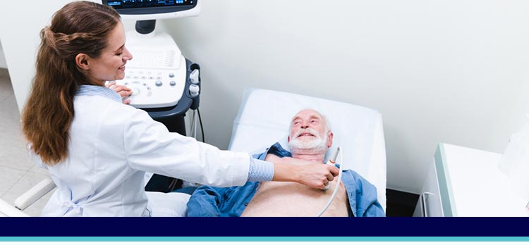
● During the Procedure: Once you’re in the examination room, you’ll lie on an exam table, and a technician will apply a special gel to your chest to help with sound wave transmission. Using a transducer, the technician will take images of your heart. The procedure is generally painless and typically lasts anywhere from 30 minutes to an hour, depending on its complexity.
Following these guidelines ensures your echocardiogram is conducted efficiently, providing your healthcare team with crucial information to assess your heart health accurately.
For family practitioners and internists, echocardiograms are invaluable in assessing patients who present with symptoms indicative of heart issues, such as chest pain, shortness of breath, or unexplained fatigue. While an echocardiogram does not detect a heart attack as it occurs, it plays a significant role in evaluating the aftermath of such events and other cardiac concerns.
In practice, an echocardiogram allows healthcare providers to:
1. Assess Heart Structure and Function: By providing detailed images of the heart, practitioners can evaluate the size and shape of the heart, the condition of its chambers and valves, and its pumping strength. This information is critical for diagnosing conditions such as heart failure, valve disorders, and cardiomyopathies.
2. Identify Damage from Past Heart Attacks: If a patient has suffered a myocardial infarction (heart attack), an echocardiogram can reveal damaged areas of heart muscle that may not be contracting properly. Identifying these areas helps in assessing the extent of cardiac damage and planning appropriate interventions.
3. Detect Complications: Echocardiograms can uncover potential complications from heart attacks, such as blood clots within the heart, valve dysfunction, or pericardial effusion (fluid around the heart). Recognizing these issues early can guide treatment decisions and improve patient outcomes.
4. Evaluate Overall Cardiac Health: Beyond diagnosing acute conditions, echocardiograms are a key part of routine cardiac assessments in patients with risk factors for heart disease, such as hypertension, diabetes, and high cholesterol.
In conjunction with other diagnostic tests like electrocardiograms (ECGs), blood tests for cardiac enzymes, and coronary angiography, echocardiograms provide a comprehensive view of a patient’s cardiac health.
An echocardiogram is a highly accurate and reliable diagnostic tool used to assess the heart’s structure and function. It uses sound waves to create detailed images of the heart, allowing healthcare providers to evaluate its chambers, valves, and blood flow. The accuracy of an echocardiogram largely depends on the type of test (such as transthoracic or transesophageal), the skill of the technician, and the quality of the equipment used. In general, echocardiograms provide valuable insights into conditions like heart valve diseases, congenital heart defects, and heart failure, making them an essential part of cardiac care.
However, like any medical test, an echocardiogram is not without its limitations. The accuracy can sometimes be influenced by factors such as the patient’s body type, lung disease, or the presence of obesity, which may hinder the ability to obtain clear images. For instance, in patients with a thick chest wall or excessive lung tissue, sound waves may not penetrate as effectively, leading to lower-quality images. In these cases, additional imaging tests may be recommended to supplement the echocardiogram results.
Despite these potential limitations, echocardiograms are considered one of the best non-invasive methods for heart evaluation. They can accurately assess heart function in most patients and are often the first-line diagnostic tool for conditions like heart murmurs, arrhythmias, or valve problems. In rare cases where an echocardiogram does not provide a definitive diagnosis, other tests, such as MRI or CT scans, might be recommended to further investigate heart health. Overall, when performed properly, echocardiograms are a reliable and essential tool in cardiology.
The frequency of echocardiograms largely depends on a person’s health status, medical history, and risk factors. For individuals with known heart conditions, such as heart valve disease or heart failure, regular echocardiograms are often part of ongoing care. These tests help monitor the progression of the condition and evaluate the effectiveness of treatments. Depending on the severity and the doctor’s recommendations, an annual or bi-annual echocardiogram may be suggested to ensure heart health is effectively managed.
For individuals with risk factors such as a family history of heart disease, hypertension, or other cardiovascular concerns, periodic echocardiograms may also be recommended. These tests serve as a proactive measure to detect potential heart problems early, even if the person does not yet show symptoms. This approach helps in identifying issues before they become more serious, allowing for timely interventions.
For those experiencing symptoms like chest pain, shortness of breath, or unexplained fatigue, an echocardiogram is essential for diagnosis. If abnormalities are detected, follow-up tests may be necessary to track the condition and make adjustments to treatment as needed. The frequency of follow-up echocardiograms will depend on the findings from the initial test and the individual’s ongoing heart health.
The decision on how frequently to schedule echocardiograms is made collaboratively between the patient and their healthcare provider. Internal Medicine and Family Practice physicians will consider the patient’s overall health, existing conditions, and risk factors during routine check-ups to devise a personalized echocardiogram schedule.
At Internal Medicine and Family Practice in Joliet, IL, Dr. Samiullah Choudry provides complete heart health insights through echocardiogram and EKG testing. These diagnostic tools reveal critical details about your heart’s rhythm, valves, and overall function. With Dr. Choudry’s expertise, patients gain a deeper understanding of their cardiovascular health and the steps necessary to protect it for years to come.
Regular appointments are important to review health status and make any necessary adjustments to the care plan, ensuring optimal heart health and overall well-being. For more information about echocardiograms, we welcome you to contact us or schedule an appointment! We are located at 1719 Glenwood Ave Joliet, IL 60435. We serve patients from Joliet IL, Plainfield IL, Lockport IL, Channahon IL, Romeoville IL, Manhattan IL and surrounding areas
Check Out Our 5 Star Reviews


